
What Is a 2D Echocardiography?
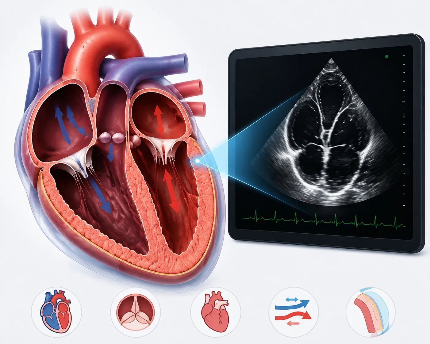
Think of it as an ultrasound, but for your heart instead of a baby bump. A technician moves a small probe called a transducer across your chest; it sends out sound waves, and those waves bounce back to build a live, moving picture of your heart on a screen. No radiation, no injections just sound.
The difference from an ECG is simple: an ECG only picks up electrical signals, so it can tell you something’s off but not what. A 2D Echo actually shows the chambers filling up, the valves snapping shut, the whole mechanism working (or not working) in front of you. Takes about twenty to thirty minutes at the clinic, start to finish.
“A 2D Echo gives us a real-time view of how the heart is structured and how it is working. When interpreted along with the patient’s symptoms and other investigations, it can help us understand the heart more clearly and decide what needs to be done next.”
Dr Krishna Dhoot, Interventional Cardiologist in Pune
Looking for a 2D Echo in Pune?
Book an appointment with Dr. Krishna Dhoot for a comprehensive cardiac evaluation.
What Is a 2D Echocardiography?
Because sometimes hearing that something’s wrong isn’t enough; you need to see it. That’s the whole point of this test. Doctors use it to check how well the heart is pumping (there’s a technical term for this, ejection fraction, you’ll hear it thrown around), to watch whether the valves open and close the way they should, and to catch structural problems, whether those have been there since birth or crept in over the years. If you’re already on treatment for a heart condition, this is also how Dr. Dhoot checks whether that treatment is actually doing its job, rather than just hoping it is.
When Is a 2D Echo Recommended?
There’s no single trigger it’s usually a combination of things.
Breathlessness that seems disproportionate to what you’re doing.
Fatigue that doesn’t go away no matter how much you rest. Swollen ankles.
Swollen ankles
A murmur someone picked up during a routine checkup.
Palpitations that won’t quit.
Recovering from a heart attack.
Blood pressure that refuses to come under control despite medication.
A family history of heart problems that makes you (rightly) a little paranoid.
What Is a 2D Echocardiography?
Quite a lot, actually, which is what makes it such a workhorse of a test. Valve problems, whether that’s narrowing or leaking. How efficiently your heart is pumping overall. An enlarged chamber, a thickened muscle wall, fluid sitting around the heart where it shouldn’t be. Congenital issues too, like a small hole between chambers that nobody noticed for years because it never caused obvious symptoms. And for anyone recovering from a cardiac event, this test shows how much muscle got affected and how well the rest of the heart is picking up the slack.
Types of Echocardiography
01
The standard version, and almost always where things start. Flat, moving images that show chamber size, wall thickness, and valve movement with enough detail to answer most questions without needing anything fancier.
02
This adds motion data. It measures how fast and in which direction blood is moving through your heart and vessels, which is useful for spotting a leaky valve or unusual flow that a plain image might not make obvious.
03
Same idea as Doppler, but the blood flow shows up in colour, layered right on top of the moving heart image. Makes it a lot easier to spot backward leakage or turbulence at a glance instead of squinting at grayscale numbers.
How Is a 2D Echo Test Performed?
You lie on your left side that position brings the heart closer to the chest wall, which just makes for a cleaner image. A bit of gel goes on your chest (it’s cold, fair warning), and the technician runs the probe over different spots while the picture builds on the monitor. No pain, maybe a bit of pressure from the probe itself. Twenty, thirty minutes and you’re done. Wipe off the gel, grab your bag, go back to your day. No recovery time, nothing to worry about after.
Ask About 2D Echo Availability
2D Echocardiography Cost in Pune
Prices bounce around depending on where you go and which version of the scan you need; colour Doppler naturally costs more than a plain 2D Echo since there’s more going on technically. At Hrudaysparsh Heart Care Centre, a standard 2D Echo is ₹2,000, and that includes Dr. Dhoot personally going through the images with you, not just handing over a printout. Compare that to getting scanned at a big hospital and then paying separately for someone to actually explain what it means it adds up fast. If your case needs Doppler or colour Doppler on top of the basic scan, that gets explained clearly before anything happens, so there’s no surprise on the bill later.
Standard 2D Echo
2,000
Includes Dr. Dhoot personally going through the images with you — not just a printout.
Why Choose Dr. Krishna Dhoot for 2D Echocardiography in Pune?
Choosing the right cardiac specialist matters because an echocardiogram is more than just an image or a printed report. The findings need to be understood in relation to your symptoms, medical history and other investigations.
Dr. Krishna Dhoot is a cardiologist in Pune with 15+ years of experience in cardiac care. He is the Director of Hrudaysparsh Heart Care Centre and provides evaluation and management for a wide range of heart conditions. His experience includes both diagnostic and interventional cardiology, with
15+
Years of experience
30,000+
Angiographies
10,000+
Angioplasties
Patients seeking a 2D Echo can benefit from:
Experienced cardiology evaluation
Patient-focused consultation
Comprehensive assessment of heart health
ECG and other cardiac diagnostic services
Guidance based on symptoms and medical history
Evaluation of abnormal or concerning cardiac findings
Appropriate recommendations for further testing when required
A 2D Echo can sometimes identify a finding that needs further attention, but an abnormal result does not automatically mean that you have a serious heart condition. Your cardiologist will explain what the findings mean and whether any additional evaluation or treatment is necessary.
that concern you, speak with Dr. Krishna Dhoot for a proper cardiac assessment.
Frequently Asked Questions
1. Does a 2D Echo hurt?
No. Maybe a little pressure from the probe as it moves around; that’s it.
2. How long does it take?
Usually twenty to thirty minutes, sometimes a touch longer if the technician needs extra angles for a clearer view.
3. Anything I need to do beforehand?
Nope. No fasting, no changing your medicines, just show up and go about your day like normal.
4. What's actually different between a 2D Echo and a Colour Doppler Echo?
The 2D shows you the structure — how the heart looks and moves. Colour Doppler adds blood flow on top of that, shown in colour, so you can see how well blood is moving through the valves.
5. Can it catch a hole in the heart?
Yes, that’s actually one of the more common things it picks up, especially congenital ones people don’t know they have.
6. Will I automatically need a Doppler scan too?
Not automatically, no. Dr. Dhoot will only add it if the basic scan suggests it’s worth looking closer.
7. How often should this be repeated?
Depends entirely on your situation — existing conditions, symptoms, risk factors. Ask Dr. Dhoot during your visit, and he’ll give you a straight answer based on your case specifically.
8. Is it safe for pregnant women or older patients?
Yes, completely. It runs on sound waves, not radiation, so age or pregnancy isn’t a concern here.